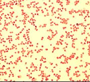
Diplocoque Gram Positif
Encyclopedie Larousse En Ligne Bacterie Latin Scientifique Bacterium Du Grec Bakterion Petit Baton

Gram Negatif Wikipedia
Medecine Paris 5
Q Tbn And9gctemtgtpwnngq0on5qwh Mf1ejuffps7xectpscp5cjgjbq6qwe Usqp Cau
Bacteriologie Des Infections Orl Infections Respiratoires Ppt Video Online Telecharger
Identification Des Cocci Gram Ppt Telecharger
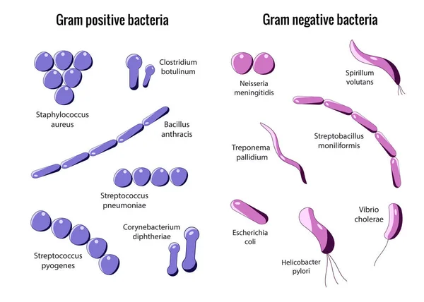
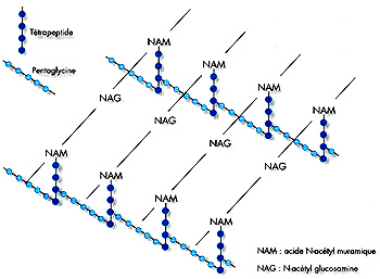
Gram positif Protéines de surface Acides Téichoiques LTA Acide Lipo téichoiques Peptidoglycane Membrane cytoplasmique Protéines Transmembranaires Paroi.
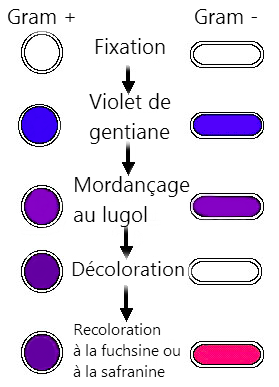
Diplocoque gram positif. J'ai mis cette vidéo espérant que sa va vous donner une idée sur les cocci a gram négatif N’oublie pas de partagé. Grampositive bacteria take up the crystal violet stain used in the test, and then appear to be purplecoloured when seen through an optical microscopeThis is because the thick peptidoglycan layer in the bacterial cell wall retains the stain after it is washed away from the rest of the sample, in the decolorization stage of the test Conversely, gramnegative bacteria cannot retain the. Diagnostic positif Clinique Signes généraux Fièvre frisson, altération de l’état général refus de tété parfois pâleur cutanéomuqueuse avec splénomégalie Syndrome respiratoire fait de gène respiratoire faite de polypnée avec des signes de lutte Toux gênante fréquente sèche puis productive.
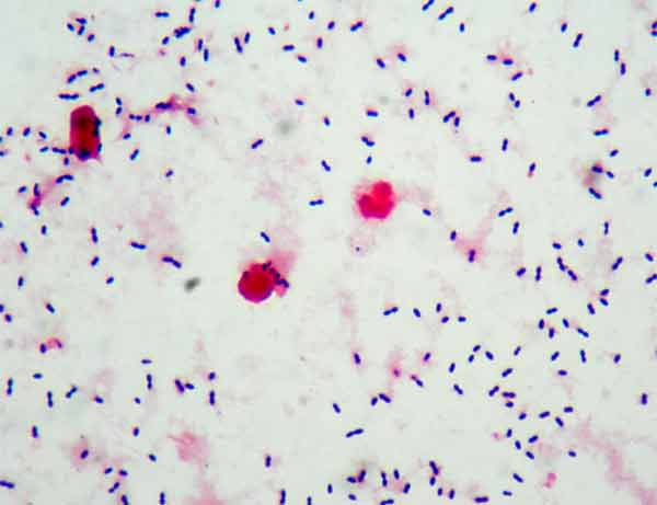
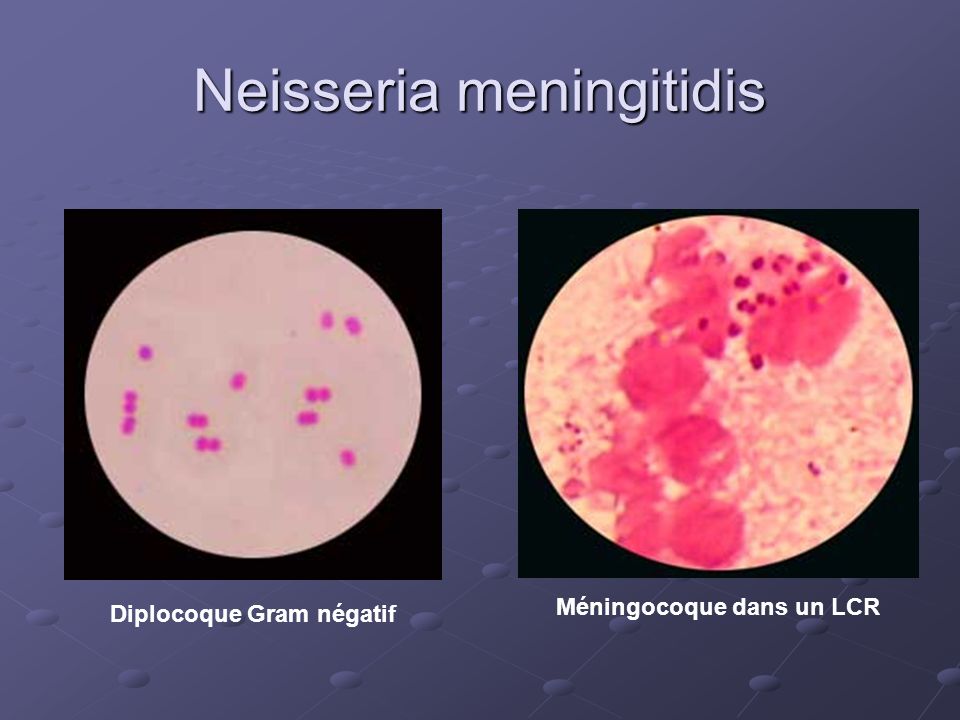
Grampositive bacteria to include methicillinresistant Staphylococcus aureus (MRSA), methicillinsusceptible Staphylococcus aureus (MSSA), and enterococci, to include vancomycinresistant enterococci (VRE), display a remarkable array of resistance and virulence factors, which have contributed to their prominent role in infections of the critically ill. Pour décrire ces grains (kokkos) groupés en amas irré. Un diplocoque Gram est ainsi isol6/~ partir des pus ur6thraux et des rhinopharynx Cette bac t6rie oxydase positive, hydrolyse glucose et mal tose, une identification pr6cise montre qu'il s'agit de m6ningocoques.
Grampositive bacteria take up the crystal violet stain used in the test, and then appear to be purplecoloured when seen through an optical microscopeThis is because the thick peptidoglycan layer in the bacterial cell wall retains the stain after it is washed away from the rest of the sample, in the decolorization stage of the test Conversely, gramnegative bacteria cannot retain the. Gram stain testing is a method for classifying bacteria based on their cell wall It allows scientists to determine whether an organism is grampositive or gramnegative The test, which uses a. CGPpr Cocci Gram Positif en Paire ;.
Klasifikasi Bakteri Gram Positif Bakteri umumnya berbentuk 1sel atau sel tunggal atau uniseluler, tidak mempunyai klorofil berkembangbiak dengan pembelahan sel atau binerKarena tidak mempunyai klorofil, bakteri hidup sebagai jasad yang saprofitik ataupun sebagai jasad yang parasitikTempat hidupnya tersebar di manamana, yaitu di udara, di dalam tanah, didalam air, pada bahanbahan, pada. Le pneumocoque est souvent retrouvé par paire de deux bactéries accolées (diplocoque) Ce sont des bactéries du type Gram positif Le génome est assez petit,. In this video, I will be discussing various mnemonics on easily remembering various Gram positive & negative cocci and bacilli.
Un bacille, c'est une bactérie Gram négatif, ça fait référence à une coloration avec une solution gram, utilisée pour "détecter" les germes ils peuvent être gram positif, ou gram négatif, en soi, ça permet juste de différencier certains germes Ce n'est pas une indication de leur pouvoir infectieux. Coloration ZN spécifique , diagnostic de la tuberculose Bactéries Gram positif et bact à. Bakteri gram positif yang dalam bahasa Inggris dituliskan “Grampositive bacteria” adalah pengelompokkan bakteri yang berdasarkan pewarnaan gram yang sebelumnya diciptakan oleh Hans Christian Gram, seorang peneliti Denmark, pada tahun 14 Silahkan dibaca tentang metodenya pada artikel dibawah ini.
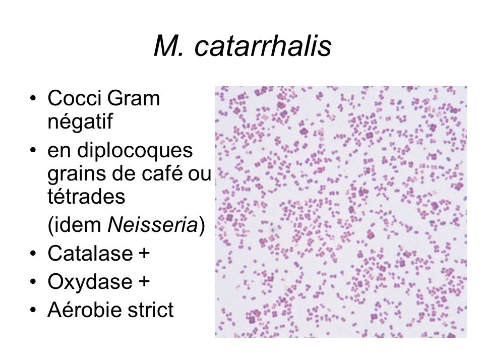
These grampositive, coccoid bacteria were once thought to be harmless to the human body However, within the last ten years, there has been an influx of nosocomial pathogens originating from Enterococcus bacteria Pathogenicity Many of these diplococci bacteria have species (strains) exhibit pathogenic characteristics. Neisseria meningitidis(Méningocoque)• Diplocoque gram négatif• 03 sérotypes (A,B,C)• Le sérotype A est fréquent en Algérie• Enfant d’âge scolaire 10 Streptococcus pneumoniae (Pneumocoque)• Cocci gram positif• Terrain particulier traumatisme crânien, intervention ORL, immunodéprimés 11. The term "gram positive" indicates that the microorganisms retain violet dye when stained using the Gram staining method Most of these organisms are susceptible to treatment with antibiotics, but several forms have developed resistance to antibiotics and require more aggressive forms of treatment An example is the potentially fatal superbug.
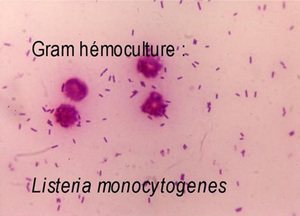
Diplocoque Gram positif = méningite à pneumocoque Diplocoque Gram négatif = méningite à méningocoque Bacille Gram positif = méningite à Listeria Only gold members can continue reading. Prokaryotes are identified as grampositive if they have a multiple layer matrix of peptidoglycan forming the cell wall Crystal violet, the primary stain of the Gram stain procedure, is readily retained and stabilized within this matrix, causing grampositive prokaryotes to appear purple under a brightfield microscope after Gram staining For many years, the retention of Gram stain was one of. In a Gram stain test, bacteria are washed with a decolorizing solution after being dyed with crystal violetOn adding a counterstain such as safranin or fuchsine after washing, Gramnegative bacteria are stained red or pink while Grampositive bacteria retain their crystal violet dye This is due to the difference in the structure of their bacterial cell wall.
Start studying Bactériologie Cours 1 Généralités Learn vocabulary, terms, and more with flashcards, games, and other study tools. A – Diplocoque Gram positif B Bâtonnet Gram Positif C Cocci en grappe, Gram négatif D Bâtonnet Gram négatif E Cocci en grappe, Gram positif F Bacille non colorable 2 Vibrio cholerae où se multiple t il plus spécialement chez l'homme?. Bactéries à Gram positif Bactéries à Gram négatif Mycobactéries Pr E Cambau 10 Classification morphologique • Cocci – à Gram positif coloration de Gram cocci à Gram positif en diplocoque Streptococcus pneumoniae ou pneumocoque Streptocoque Pr E Cambau 13 Examen microscopique après.
De Médecine de Constantine Cours de Microbiologie Pr HLaouar Bactéries à. Bakteri Gram Positif Mesosom lebih menonjol pada bakteri gram positif Bakteri Negatif Gram Mesosom kurang menonjol pada bakteri gram negatif Ketahanan terhadap Gangguan Fisik, Sodium Azide, dan Pengeringan Bakteri Gram Positif Resistensi terhadap gangguan fisik, natrium azida, dan pengeringan tinggi bakteri gram positif. CVD Center for Vaccine Development ;.
Le Streptococcus pneumoniae est un diplocoque, Gram positif dont toutes les souches sauvages possèdent une capsule composée de polysaccharides agissant comme une armure. A) MORPHOLOGIE Elle est très variable selon le G , parfois l’espèce considérée Exemples G Finegoldia Emagna Cocci a Gram en diplocoque, tetrades et chainettes, immobile, peut etre capsulé GActinomyces israelii;. فطرة اصبح البول عندي لونه احمر عملت تحليل بول وظهر عندي diplocoque gram positif G وبعدها ظهرت بقع حمراء بين القضيب والخصية وختفت وعندما بدات بالعلاج ظهرت بقعة واحدة حمراء في راس القضيب وبعد ثلاثة ايام.
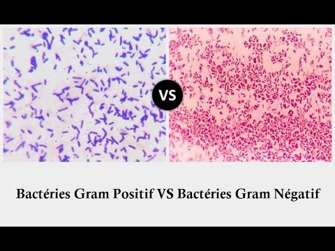
Streptococcus pneumoniae *Diplocoque Gram (), encapsulé *Saprophyte des VR hautes *Germe de transmission interhumaine *Colonise enfant dès 1ers jours de vie *Forme de « 8 » ou « gousse d’arachide » *Clinique cf TDD Staphylocoques Cocci Gram positif Anaérobies Terrains débilité, DDB, cancer toxicomanie IV, cathétérisme. When the density of vancomycinresistant enterococci in stool was at least 4 log per gram, 10 of 12 sets of cultures of environmental specimens had at least one positive sample, as compared with 1. Les bactéries à Gram positif sont mises en évidence par une technique de coloration appelée coloration de GramCette technique de coloration, qui permet de classifier les bactéries dans deux catégories générales, repose sur les caractéristiques membranaires et de paroi de la bactérie Les bactéries à Gram positif apparaissent alors mauves au microscope.
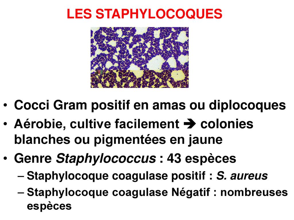
Bactéries à Gram positif A/Les staphylocoques IIntroduction Les staphylocoquesont été découverts dans un pus par Pasteur en 10 En 18 Ogston a crée le nom de « Staphylocoque » pour décrire ces grains (kokkos) groupés en amas irréguliers a la façon d'une grappe de raisin (staphylos) En 14, Rosenbach a obtenu des. Bacilles a Gram, rassemblés en forme de branches a ramifications courtes, peu abondantes. Bakteri gram positif akan lebih mempertahankan warna dasar karena dinding selnya yang tebal dan menyerap warna lebih banyak sehingga ketika dilakukan dekolorisasi, maka warna dasar tetap bertahan Berbeda dengan bakteri gram negatif yang hanya memiliki dinding sel selapis, warna dasar yang diberikan akan dengan mudah luntur atau tercuci saat.
The perbezaan utama antara bakteria gram positif dan gram adalah bahawa bakteria gram positif mengandungi dinding sel peptidoglycan yang tebal bersamasama dengan asid teikoid, membenarkan bakteria menjadi noda dalam ungu semasa pewarnaan gram manakala bakteria gram negatif mengandungi dinding sel peptidoglycan nipis tanpa asid teikoik. Gram Positive Bacteria The cell walls of Gram positive bacteria differ structurally from the cell walls of Gram negative bacteria The primary component of bacterial cell walls is peptidoglycan Peptidoglycan is a macromolecule composed of sugars and amino acids that are assembled structurally like woven material The amino sugar component consists of alternating molecules of N. Bacilles à Gram négatif, origine hydrotellurique Contamination par aérosols d’eau contaminée Déclaration obligatoireévidence par des colonies (halo vert sur gélose au sang) Diagnostic ou respiratoire Production de pneumolysine mise en αhémolytiques Diplocoque à Gram positif Genre Streptococcus avec capsule polyosidique.
Grampositive cocci are included among some of the most significant human bacterial pathogens primary pathogens such as Staphylococcus aureus, Streptococcus pyogenes, and Strep pneumoniae, along with species of lower virulence such as Staph epidermidis, Staph saprophyticus and Enterococcus faecalis Isolation and identification of these organisms is one of the most important but also routine tasks performed in clinical microbiology. Bakteri Gram Positif Berwarna Ungu Bakteri Gram positif mengandung ikatan lapisan peptidoglikan yang tebal sehingga kuat mengikat pewarna primer berupa Kristal Violet, setelah diikuti penambahan larutan mordant berupa lugols Iodine Kristal Violet dan Lugols Iodine akan membentuk ikatan komplek dengan peptidoglikan. DCGN Diplocoque Gram Négatif ;.
Le Streptococcus pneumoniae, ou plus communément le pneumocoque, est un diplocoque, Gram positif Toutes les souches sauvages de S pneumoniae possède une capsule composée de polysaccharides qui agit comme une armure contre le système immunitaire de l’hôte infecté. Grampositif adalah bakteri yang mempertahankan zat warna kristal violet sewaktu proses pewarnaan Gram sehingga akan berwarna biru atau ungu di bawah mikroskop Disisi lain, bakteri gramnegatif akan berwarna merah atau merah muda Perbedaan keduanya didasarkan pada perbedaan struktur dinding sel yang berbeda dan dapat dinyatakan oleh prosedur pewarnaan Gram. Streptococcus pneumoniae are grampositive, lancetshaped, polysaccharideencapsulated diplococcus It frequently colonizes the upper respiratory tract and may cause infection or invasive disease It has a high morbidity in children, especially when related with the respiratory tract.
Demonstration of gramnegative dipplococci or signs associated to CSF aspect of bacterial meningitis (petechiae, positive PCR or detection of N meningitidis antigens), was found for 5 patients (78%) Among those cases, meningococcemia characterized by isolation of N meningitidis, positive. Demonstration of gramnegative dipplococci or signs associated to CSF aspect of bacterial meningitis (petechiae, positive PCR or detection of N meningitidis antigens), was found for 5 patients (78%) Among those cases, meningococcemia characterized by isolation of N meningitidis, positive. Resistance to fluoroquinolones among Grampositive cocci has emerged as these antimicrobial agents have become extensively used in clinical medicine Resistance is effected by changes in the bacterial target enzymes DNA gyrase and topoisomerase IV, which reduce drug binding, and by action of native.
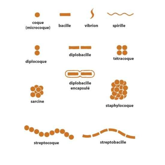
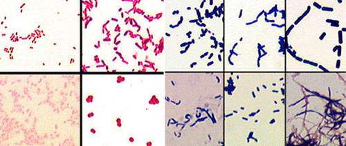
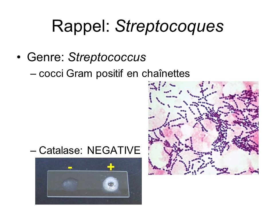
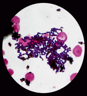
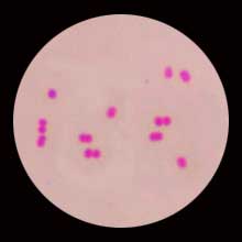
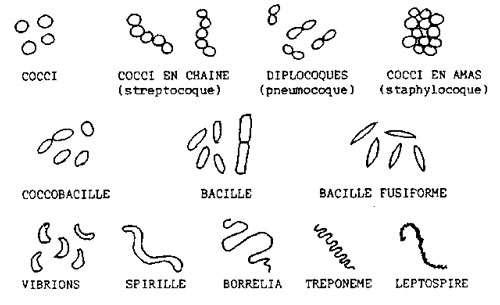
A – Diplocoque Gram positif B Bâtonnet Gram Positif C Cocci en grappe, Gram négatif D Bâtonnet Gram négatif E Cocci en grappe, Gram positif F Bacille non colorable 2 Vibrio cholerae où se multiple t il plus spécialement chez l'homme?. Bactéries à Gram positif A/Les staphylocoques I Introduction Les staphylocoques ont été découverts dans un pus par Pasteur en 10 en 18 Ogston a crée le nom de « Staphylocoque » pour décrire ces grains (kokkos) groupés en amas irréguliers a la façon d'une grappe de raisin (staphylos) En 14, Rosenbach a obtenu des cultures pures. Diplocoque Ex Streptococcus pneumoniae Chaines de cocci Ex Streptococcus mutans Chaines de cocci Ex Streptococcus mutans Grappe Ex staphylococcus aureus Packet Ex Streptococcus pyogenes Paroi des Gram positif ultrastructure Deux couches dont l’une dense et epaisse 38.
Chart and Diagram Slides for PowerPoint Beautifully designed chart and diagram s for PowerPoint with visually stunning graphics and animation effects Our new CrystalGraphics Chart and Diagram Slides for PowerPoint is a collection of over 1000 impressively designed datadriven chart and editable diagram s guaranteed to impress any audience. Gram négatif Paroi des bactéries à. فطرة اصبح البول عندي لونه احمر عملت تحليل بول وظهر عندي diplocoque gram positif G وبعدها ظهرت بقع حمراء بين القضيب والخصية وختفت وعندما بدات بالعلاج ظهرت بقعة واحدة حمراء في راس القضيب وبعد ثلاثة ايام.
Gram positif A/Les staphylocoques I Introduction Les staphylocoques ont été. Sample patients with IE caused by grampositive cocci, having received at least 10 days of conventional antibiotic treatment, and at least 7 days after surgery when indicated, without clinical, analytical, microbiological or echocardiographic signs of persistent infection Estimated sample size 298 patients. Introduction Le Streptococcus pneumoniae est un diplocoque, Gram positif dont toutes les souches sauvages possèdent une capsule composée de polysaccharides agissant comme une armure protectrice contre le système immunitaire de l’hôte infecté Pour la confectionner, la bactérie utilise 92 polysaccharides dont la structure et le motif antigénique sont différents qui sont classés en.
A dans la lumière intestinale B dans l'estomac C dans les voies biliaires D dans le. Diplocoque Gram ressemblant beaucoup à Neisseria meningitidis (mais non capsulée) Elle a un réservoir strictement humain, et est la 2ème cause d'urétrite après le Chlamydia trachomatis Chez l'homme urétrite aiguë, épididymite et prostatite, chez la femme urétrite et cervicite passant souvent inaperçu. GRAMPOSITIF adalah bakteri yang mempertahankan zat warna kristal violet sewaktu proses pewarnaan Gram sehingga akan berwarna biru atau ungu di bawah mikroskop Disisi lain, bakteri gramnegatif akan berwarna merah atau merah mudaPerbedaan keduanya didasarkan pada perbedaan struktur dinding sel yang berbeda dan dapat dinyatakan oleh prosedur.
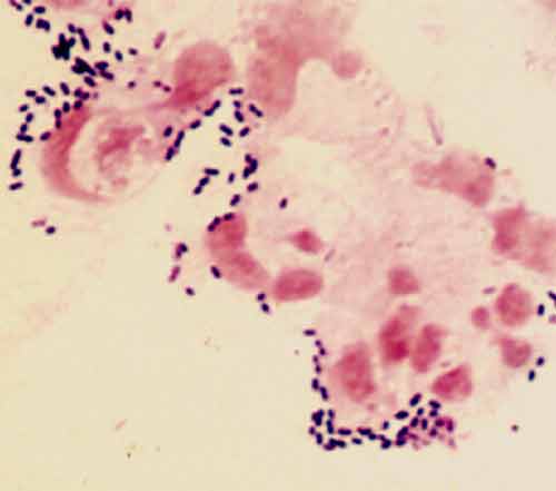
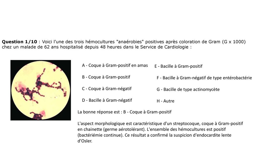
Pengertian pewarnaan gram Pewarnaan gram atau disebut juga metode gram merupakan metode pewarnaan yang biasa digunakan untuk membedakan dan mengklasifikasikan spesies bakteri menjadi dua kelompok besar yaitu bakteri gram positif dan bakteri gram negatif Bakteri gram positif memiliki peptidoglikan yang tebal (90% dari dinding sel) dan berwarna ungu saat diwarnai dengan pewarnaan gram. A dans la lumière intestinale B dans l'estomac C dans les voies biliaires D dans le. Les diplocoques sont des bactéries que l'on retrouve sous forme de petits grains de café identiques accolées deux à deux à l'observation au microscope 1 Ces agents microbiens sont caractérisés par leur potentiel infectieux Les infections par le Méningocoque (gram ), le Gonocoque (gram ) ou encore le Pneumocoque (gram ) sont caractérisées par cette forme de regroupement.
Pengertian Bakteri Gram Positif Bakteri gram positif adalah bakteri yang dinding selnya menyerap warna violet dan memiliki lapisan peptidoglika yang tebal Bakteri gram positif memiliki ciriciri sebagai berikut Dinding sel bersifat homogen dan memiliki ketebalan 80 nm. Cocci Gram Positif en Grappe ;. Bacilles a Gram, rassemblés en forme de branches a ramifications courtes, peu abondantes.
The term "gram positive" indicates that the microorganisms retain violet dye when stained using the Gram staining method Most of these organisms are susceptible to treatment with antibiotics, but several forms have developed resistance to antibiotics and require more aggressive forms of treatment An example is the potentially fatal superbug. Découverts dans un pus par Pasteur en 10 en 18 Ogston a crée le nom de «. Prokaryotes are identified as grampositive if they have a multiple layer matrix of peptidoglycan forming the cell wall Crystal violet, the primary stain of the Gram stain procedure, is readily retained and stabilized within this matrix, causing grampositive prokaryotes to appear purple under a brightfield microscope after Gram staining For many years, the retention of Gram stain was one of.
A) MORPHOLOGIE Elle est très variable selon le G , parfois l’espèce considérée Exemples G Finegoldia Emagna Cocci a Gram en diplocoque, tetrades et chainettes, immobile, peut etre capsulé GActinomyces israelii;. 1b Coloration de Gram Présence de cocci en diplocoque à Gram positif intracellulaire Les forme s extracellulaires sont rencontrées vu leur éclatement par la technique de coloration 2 Gonococcie chronique Le gonocoque se raréfie et sa recherche doit se faire très longtemps vue l'absence de la réaction cellulaire.
Http Www Umft Ro Data Files Documente Atasate Sectiuni 5726 Manuel de microbiologie 2 Pdf

Bacteries Gram Positives Vs Gram Negatives
Www Studocu Com Fr Document Universite Paris Saclay Bacteriologie Notes De Cours Fiches De Cours Ue 3b Bacteriologie View
Pharmacie Ma Uploads Pdfs Le Guide Pratique Des Bacteries Pathogenes Pdf

Streptococcus Pneumoniae Wikipedia
Http Univ Ency Education Com Uploads 1 3 1 0 Bacterio3an Gram Positif17 Laouar Pdf
Identification Bacterienne Par La Coloration De Gram Biotechnologie
Les Differentes Bacteries Gram Et Sa Coloration Dossier
Medecine Paris 5
Spiral Cours Bacteriologie Des Angines
Http Wd Fmpm Uca Ma Biblio Epreuves Epreuves Reservoire 17 12 Pdf

Cours

Coques Gram Catalase Moins
Www Eurofins Biomnis Com Referentiel Liendoc Precis Pneumococcie Pdf
2

Qu Est Ce Qu Une Bacterie
Cocci Gram Positif

Prelevements Genitaux
Les Differents Types De Bacteries Les Antibiotiques
Cours Examens Org Images An 18 Etudes Superieures Biologie Bacteriologie Autres 15 Pdf
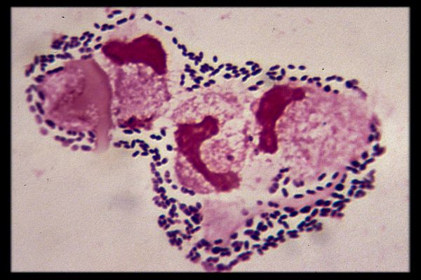
Liquide Cephalo Rachidien Examens Directs Ou Portraits Des Bacteries
Syndrome Meninge Syndrome Septicemique Ppt Video Online Telecharger
Pharmacie Ma Uploads Pdfs Le Guide Pratique Des Bacteries Pathogenes Pdf

Hemocultures Avec Bacteries A Gram Positif Portraits Des Bacteries Ou L Examen Direct

Developpement Et Sante Colorations Usuelles En Bacteriologie
Www Chu Nantes Fr Medias Fichier Antibiotherapie Ifsi 18 Pdf

The In Vitro Aid Response To The Vaccine Tracks With The Serum Response Download Table

Hemocultures Avec Bacteries A Gram Positif Portraits Des Bacteries Ou L Examen Direct
Cocci Gram Positif
Q Tbn And9gcrjep5hscrgopsrcbktltrzsvdjzknd4pcj2gx6wcxezfmaypbd Usqp Cau

Cocci Wikipedia

Bacteries Gram Positives Vs Gram Negatives
Http Wd Fmpm Uca Ma Biblio Epreuves Epreuves Reservoire 15 12 Pdf
Examen Direct Bacteriologie Ppt Telecharger

Cocci Gram Positif Staphylococcus Streptocoque

Espace Techniciens De Laboratoire Les Coccis Gram Positif Staphylococcus Aureus

Examen Direct Bacilles A Gram Negatif En Diplocoque D Aspect Polymorphe Download Scientific Diagram

La Coloration De Gram L Exsudat Uretral Des Hommes Avec L Uretrite 1974 Remarque L Absence De Diplocoques Intracellulaires Gram Negatif La Technique De Coloration De Gram Qui Emploie Crystal Violet Comme La Tache Principale
Bacteriologie Des Infections Orl Infections Respiratoires Ppt Video Online Telecharger

Qcm D Aujourd Hui Home Facebook
Http Wd Fmpm Uca Ma Biblio Epreuves Epreuves Reservoire 15 12 Pdf
Les Bacteries Gram Positif Et Gram Negatif Youtube
Http Www Ifsidijon Info V2 Wp Content Uploads 17 09 Ifsiagentsinfectieux17 Pdf

Medecine Paris 5
Www Ctcb Com Documentation Fiches techniques bac Rothia mucilaginosa Edition 10 Pdf
3 Gram Confusions
Bacteries Gram Positives Vs Gram Negatives

Gram Positif Wikipedia

Diplocoques Banque D Image Et Photos Alamy
Examen Direct Bacteriologie Ppt Telecharger
Http Www Infectiologie Com Userfiles File Medias Jni Jni08 Inf Syndmening ide08 Gouriet Pdf

Identification D Acinetobacter Spp Au Laboratoire Sciencedirect
Gram Stained Photomicrograph Banque D Image Et Photos Alamy
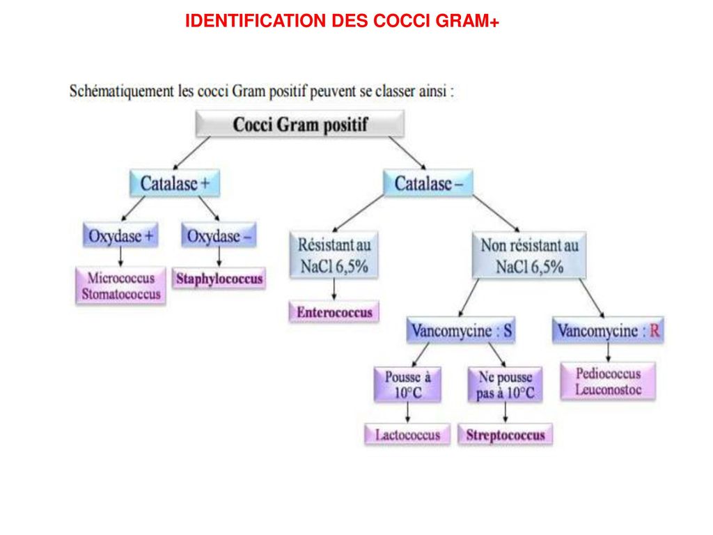
Identification Des Cocci Gram Ppt Telecharger
Q Tbn And9gcqqqg J8ik1p3c1boi4sbowb4rq4egi2acyuutrgppsltl4 Fbo Usqp Cau
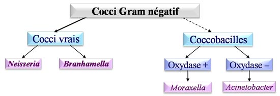
Cocci Gram Negatif

Diagnostic Des Meningites Au Laboratoire

Bacteries Et Infections Orl Ppt Video Online Telecharger
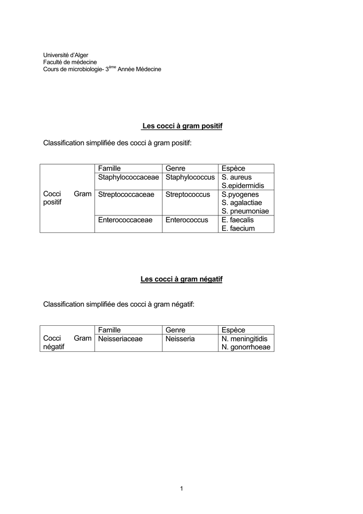
Les Cocci A Gram Positif Classification Simplifiee Des Cocci A Gram

Ppt Les Streptocoques Powerpoint Presentation Free Download Id
Site De Microbiologie Medicale
Http Www Cours Examens Org Images An 18 Etudes Superieures Biologie Bacteriologie Bacteriologie Bacteries Gouriet Pdf
Http Www Memobio Fr Formation 1 Cgp Reponses Pdf
Bacteries Anaerobies Stricts
Photos De Gram Positif Images De Gram Positif Depositphotos

Cocci Gram Positif Staphylococcus Streptocoque
Www Wiv Isp Be Qml Activities External Quality Rapports Down Microbiologie 12 12 2f Microbio Pdf
Streptocoques Enterocoques

Diplococci Dans Le Frottis Pour Les Femmes Et Les Hommes Causes Traitement Avec Competence Sur La Sante Sur Ilive

Examen Cytobacteriologique Des Crachats Coloration De Gram 100 Download Scientific Diagram

Cocci Wikipedia
Http Www Ipubli Inserm Fr Bitstream Handle 8 3fsequence 3d8
Q Tbn And9gcqbbcyvjwsmvfdszddzor3zpanj0bmqak8sykf2pjn0oqlixtdk Usqp Cau
3 Gram Confusions

Neisseria Meningitidis Wikipedia

Enterocoque Wikipedia
Pharmacie Ma Uploads Pdfs Le Guide Pratique Des Bacteries Pathogenes Pdf
Http Static Probioqual Com Pdf Doc Epu 21h00 Pdf
3 Gram Confusions
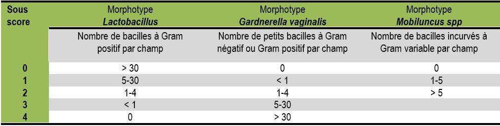
Diagnostic Des Infections Vaginales Au Laboratoire De Biologie Medicale

Hemocultures Avec Bacteries A Gram Positif Portraits Des Bacteries Ou L Examen Direct
Www Antibioest Org Wp Content Uploads 19 05 Bact C3 riemies Dur C3 e De Ttt Atb J Ref19acharmillon Pdf
Cocci A Grammes Positif Ppt Telecharger
Les Differents Types De Bacteries Les Antibiotiques
Les Differentes Bacteries Gram Et Sa Coloration Dossier

Salilab Enterocoques Diplocoques A Gram Positif Facebook

Cocci A Gram Positif Lorraine Evoluence
Www Chu Nantes Fr Medias Fichier Antibiotherapie Ifsi 18 Pdf

Qu Est Ce Qu Une Bacterie

Identification D Acinetobacter Spp Au Laboratoire Sciencedirect

Cocci A Gram Positif Dans La Chaine Banque D Images Et Photos Libres De Droits Image

Streptococcus Pneumonie Pneumocoque
Morphologie
Http Wd Fmpm Uca Ma Biblio Epreuves Epreuves Reservoire 17 12 Pdf
Http Ao Um5 Ac Ma Xmlui Bitstream Handle 176 M Pdf Sequence 1 Isallowed Y
Www Infectiologie Com Userfiles File Medias Enseignement Gericco 12 12 Gericco Cady Pdf
S4 Bacterio Cocci Gram Positif Fait Studocu
Http Www Memobio Fr Formation 1 Cgp Reponses Pdf


